DERMAGIC EXPRESS / Dermatologia y Bibliografia - Dermatology & bibliography DERMAGIC EXPRESS / Dermatologia y Bibliografia - Dermatology & bibliography

sábado, 4 de octubre de 2025

SCLEROSING LYMPHANGITIS OF THE PENIS / LINFANGITIS ESCLEROSANTE DEL PENE.


 SCLEROSING LYMPHANGITIS OF THE PENIS


LINFANGITIS ESCLEROSANTE DEL PENE


Sclerosing lymphangitis of the penis


  EDITORIAL ESPAÑOL

=================== 

Hola amigos DERMAGIC de nuevo con ustedes, el tema de hoy la LINFANGITIS ESCLEROSANTE DEL PENE. Esta patología realmente es POCO CONOCIDA, por muchos y por ello la presentamos  hoy.

1.) HISTORIA:
 
La LINFANGITIS ESCLEROSANTE DEL PENE., fue descrita por primera vez por Hoffman en 1923, quien inicialmente la denominó LINFANGITIS PLÁSTICA NO VENÉREA DEL SURCO CORONAL.
 
Se le conoce también o ha sido descrita con otros nombres:
 
A.- LINFANGITIS NO VENÉREA BENIGNA TRANSITORIA DEL PENE.
 
B.- LINFANGIO FIBROSIS OCLUSIVA TROMBÓTICA DEL PENE.
 
C.- ENFERMEDAD O FLEBITIS DE MONDOR DEL PENE. 
 
Hay que aclarar aca que la FLEBITIS DE MONDOR CLÁSICA afecta principalmente las venas subcutáneas de la pared toracoabdominal, pero como también afecta a las venas superficiales del surco balanoprepucial, se le atribuye este nombre.
 
Es una patología dermatológica poco frecuente, con pocos casos descritos en la literatura. 
 

2.) ETIOLOGÍA: 
 
A.- La hipótesis más aceptada hoy día, son MICROTRAUMATISMOS producidos por una ACTIVIDAD SEXUAL INTENSA, o la MASTURBACION, estos serían los desencadenantes principales. 

Pero si el TRAUMATISMO es la causa única, porque existen tan pocos casos descritos en la literatura ?

B.- En casos aislados se ha asociado a infecciones de transmisión sexual, como lo son el HERPES SIMPLE, CLAMIDIA, LINFOGRANULOMA VENÉREO, URETRITIS GONOCÓCICA, GRANULOMA INGUINAL y SÍFILIS.

C.- Y también se han propuesto otro tipo de etiologías NO VENÉREAS COMO: ENTEROVIRUS, TUBERCULOSIS,  o IRRITACIÓN por el sangrado menstrual de la mujer durante el acto sexual.

D.- OTROS autores la relacionan a antígenos relacionados con el FACTOR VIII de la coagulación y proponen el nombre de FLEBITIS DE MONDOR DEL PENE.

Pero las causas más aceptada sigue siendo los MICROTRAUMATISMOS.

3.) FISIOPATOLÓGICAMENTE: 

Se describe una obstrucción o inflamación de los vasos linfáticos superficiales del pene, lo que genera el cordón o engrosamiento palpable. 

El diagnóstico es clínico y debe diferenciarse de otras causas de lesiones del pene, especialmente infecciones de transmisión sexual (ETS).

4.) CARACTERÍSTICAS CLÍNICAS:

Se presenta clásicamente como como una tumefacción o inflamación lineal en forma de cordón duro y delgado, poco dolorosa, que puede rodear parcial o completamente el glande, pero principalmente se localiza en el surco coronal, es decir el surco balanoprepucial.

La lesión por lo general es indolora, pero puede causar molestias durante el acto sexual y la erección, y se nota a simple vista porque el cordón es translúcido o blanquecino, y móvil al tacto; también puede presentarse inflamación leve del prepucio o glande; en algunos casos se ha presentado erosion y ulceraciones.

Esta enfermedad afecta principalmente a hombres jóvenes de entre 18 y 45 años, con una máxima de 50 años. La lesión es benigna, por lo general, desaparece espontáneamente en varias semanas, y más rápidamente si se instaura un tratamiento adecuado.


5.) TRATAMIENTO:

A.- Reposo de la actividad sexual para evitar mayores traumatismos.

B.- Antiinflamatorios no esteroideos (AINES) si hay síntomas de dolor, o corticoides más potentes para disminuir la inflamación, por cortos periodo de tiempo.

C.- Localmente cremas emolientes y cremas con esteroides de baja potencia.

D.- Explicarle al paciente la probable causa para tranquilizarlo; pero si hay infección, diagnosticar la causa y tratarla específicamente.

El periodo de curación oscila entre 3 y 8 semanas.

6.) RESUMEN:

1.) No es una enfermedad venérea.

2.) No está causada directamente por infecciones sexuales.

3.) Es una patología rara y benigna, ocasionada por TRAUMATISMOS locales, en la mayoría de los casos.

4.) Rutinariamente debe descartarse Enfermedad de Transmisión Sexual (ETS).

Saludos a todos... 
 
Dr. Jose M. Lapenta.


linfangitis esclerosante del pene


EDITORIAL ENGLISH:
=================== 
Hello, Dermagic friends, back with you today. Today's topic is SCLEROSING LYMPHANGITIS OF THE PENIS. This condition is truly little known by many, which is why we are presenting it today.

1.) HISTORY:

SCLEROSING LYMPHANGITIS OF THE PENIS was first described by HOFFMAN in 1923, who initially called it Non-venereal lymphangitis of the coronal sulcus.

It is also known or has been described under other names:

A.) TRANSIENT BENIGN NON-VENEREAL LYMPHANGITIS OF THE PENIS.

B.) THROMBOTIC OCCLUSIVE LYMPHANGIOFIBROSIS OF THE PENIS.

C.) DISEASE OR  PHLEBITIS OF MONDOR of THE PENIS.
 
It should be clarified here that
CLASSIC MONDORS' PHLEBITIS primarily affects the subcutaneous veins of the thoracoabdominal wall, but since it also affects the superficial veins of the balanopreputial sulcus, this name is attributed to it.

It is a rare dermatological condition, with few cases described in the literature.

2.) ETIOLOGY:

A.- The most widely accepted hypothesis today is that MICROTRAUMA caused by INTENSE SEXUAL ACTIVITY or MASTURBATION would be the main triggers.

But if TRAUMA is the sole cause, why are there so few cases described in the literature?

B.- In isolated cases, it has been associated with sexually transmitted infections, such as HERPES SIMPLEX, CHLAMYDIA, LYMPHOGRANULOMA VENEREUM, GONOCOCCAL URETHRITIS, GRANULOMA INGUINAL, and SYPHILIS.

C.- Other non-venereal etiologies have also been proposed, such as ENTEROVIRUS, TUBERCULOSIS, or IRRITATION due to a woman's menstrual bleeding during sexual intercourse.

D.- Other authors associate it with antigens related to coagulation FACTOR VIII and propose the name MONDOR'S PHLEBITIS OF THE PENIS.

However, the most widely accepted cause remains
MICROTRAUMA.

3.) PATHOPHYSIOLOGICALLY:

It describes an obstruction or inflammation of the superficial lymphatic vessels of the penis, which generates the palpable cord or thickening.

The diagnosis is clinical and must be differentiated from other causes of penile injuries, especially sexually transmitted infections (STDs).

4.) CLINICAL CHARACTERISTICS:

It classically presents as a linear swelling or inflammation in the form of a hard, thin cord, with minimal pain, which may partially or completely surround the glans penis, but is mainly located in the coronal sulcus, i.e., the balanopreputial sulcus.

The lesion is usually painless, but it can cause discomfort during sexual intercourse and erection. It is noticeable to the naked eye because the cord is translucent or whitish and mobile to the touch. Mild inflammation of the foreskin or glans may also occur; In some cases, erosion and ulcerations have occurred.


This disease primarily affects young men between 18 and 45 years of age, with a maximum age of 50. The lesion is benign and usually resolves spontaneously within several weeks, and more quickly if appropriate treatment is initiated.

5.) TREATMENT:

A.- Rest from sexual activity to avoid further trauma.

B.- Nonsteroidal anti-inflammatory drugs (NSAIDs) if there are symptoms of pain, or stronger corticosteroids to reduce inflammation for a short period of time.

C.- Local emollient creams and low-potency steroid creams.

D.- Explain the probable cause to the patient to reassure them; however, if there is an infection, diagnose the cause and treat it specifically.


6.) SUMMARY:

1.) It is not a venereal disease.

2.) It is not directly caused by sexually transmitted infections.

3.) It is a rare and benign disease, caused by local trauma in most cases.

4.) Sexually Transmitted Diseases (STDs) should be routinely ruled out.

The healing period ranges from 3 to 8 weeks.


Greetings to all...

Dr. José Lapenta.

Dr. José M. Lapenta.


 ============================================================
 REFERENCIAS BIBLIOGRÁFICAS / BIBLIOGRAPHICAL REFERENCES 
 ============================================================
 ============================================================ 
 0.) SCLEROSING LYMPHANGITIS OF THE PENIS. THE HISTORY
 1.) Non-venereal sclerosing lymphangitis of the penis following herpes progenitalis.
 2.) Nonvenereal sclerosing lymphangitis of the penis.

3.) [Sclerosing lymphangitis of the penis, the upper lip and the labium minus]
4.) Nonvenereal sclerosing lymphangitis of the penis.
5.) Non-venereal sclerosing lymphangitis of the penis.
6.) Non-venereal sclerosing lymphangitis of the penis. A clinicopathologic treatise.
7.) Mondor's phlebitis of the penis. A condition miscalled 'non-venereal sclerosing lymphangitis'.
8.) Circular indurated lymphangitis of the penis.
9.) [Indications for corticotherapy in phlebology]
10.) [A case for diagnosis: sclerosing lymphangitis of the penis].
11.) [Non-venereal sclerosing lymphangitis of the penis].
12.) [Non-venereal sclerosing lymphangitis of the penis].
13.) Nonvenereal sclerosing lymphangitis of the penis.
14.) [A case of non-venereal sclerosing lymphangitis of the penis].
15.) [A case of Mondor's disease of the penis].
16.) Non-venereal sclerosing lymphangitis of the penis associated with masturbation.
17.) [2 cases of sclerosing lymphangitis of the sex organs].
18.) Mondor's phlebitis of the penis: a study with factor VIII related antigen.
19.) [Non-venereal sclerosing lymphangitis of the penis revealed as phlebitis by positive factor
VIII-related antigen].
20.) Surgical management of persistent, symptomatic nonvenereal sclerosing lymphangitis of the penis.
21.) Sclerosing lymphangitis of the penis: a lymphangiofibrosis thrombotica occlusiva.
22.) Sclerosing lymphangitis of the penis: a possible chlamydia aetiology.
23.) [Development of non-venereal sclerosing lymphangitis of the penis following herpetic balanitis].
24.) [Case of sclerosing lymphangitis of the sex organs].
25.) [Nonveneral sclerosing lymphangitis of the penis].
26.) Benign transient lymphangiectasis (sclerosing lymphangitis) of the penis.
27.) [Sclerosing lymphagitis of the penis].
28.) Lymphocoele and localized lymphoedema of the penis.
29.) Nonvenereal sclerosing lymphangitis of the penis.
30.) Sclerosing non-venereal lymphangitis of the penis].
31.) Sclerosing lymphangitis of the penis.
32.) Nonvenereal sclerosing lymphangitis of the penis.
33.) Nonvenereal sclerosing lymphangitis of the penis.
34.)The so-called Hoffman's lymphangitis of the penis: is it a lymphangitis or a phlebitis?
35.) Sclerosing lymphangitis of penis - literature review and report of 2 cases.
36.) Sclerosing lymphangitis of the penis after coadministration of tadalafil and fluconazole.
37.) [Mondor's disease vs. sclerosing lymphangitis of the penis].
38.) Immunohistochemical clues to the diagnosis of Mondor's disease of the penis.
39.) [Non venereal sclerosing lymphangitis of the penis. Case report].
40.) Mondor's disease of the penis associated with primary syphilis.
41.) Mondor's disease of penis: a forgotten disease.
============================================================ 

Si Te ha gustado, Compartelo

domingo, 21 de septiembre de 2025

EL LIQUEN PLANO, REVISIÓN. / THE LICHEN PLANUS, A REVIEW.


BUSCANDO EL ORIGEN DEL LIQUEN PLANO !!

 LOOKING FOR THE ORIGIN OF LICHEN PLANUS !!


Liquen plano cuerpo


PUBLICADO 2.017 ACTUALIZADO 2.025


EDITORIAL ESPAÑOL
===================
Hola amigos de la red. DERMAGIC de nuevo con ustedes. En esta nueva revisión sobre el LIQUEN PLANO, enfermedad descrita hace más de 150 años y que hoy día su tratamiento sigue siendo todo un reto para los dermatólogos de la nueva era.  

 
Liquen plano dorso de mano

HISTORIA:

ERASMUS WILSON fue el primero en utilizar el termino LIQUEN PLANO en el año 1869. La primera variante de la enfermedad fue descrita por Kaposi en 1892 denominándola LIQUEN RUBER PENFIGOIDES,  y frederic WICKHAM en 1895 describió las estrías blancas en la parte superior de las lesiones.
 
Posteriormente Darier en 1905 describió las características histopatológicas de la enfermedad. 
 

Después de muchos años el avance de la ciencia a logrado descifrar numerosos eventos acerca del LIQUEN PLANO, ellos son: 
 
 
ETIOLOGÍA: 
 

1.- INMUNOMEDIACIÓN 

El liquen plano es una enfermedad inflamatoria crónica que hoy dia es considerada de etiología desconocida, la cual es producida por una REACCIÓN INMUNITARIA, mediada por linfocitos T citotóxicos que atacan queratinocitos basales, causando daño y apoptosis en la piel y mucosas,  

2.) FACTORES DESENCADENANTES:
Diversos factores pueden actuar como desencadenantes, entre ellos figuran:

A.- INFECCIONES VIRALES: siendo el virus de la hepatitis C y hepatitis B las asociaciones más importantes.

B.- MEDICAMENTOS:

- Antihipertensivos: inhibidores de la enzima convertidora de angiotensina (IECA=Enalapril), y betabloqueadores: metoprolol, atenolol, propranolol, bisoprolol, carvedilol, labetalol, nebivolol).

- Aantimaláricos (cloroquina e hidroxicloroqiina). 

- Antiinflamatorios NO ESTEROIDEOS (AINES): como el ibuprofeno y naproxeno, y otros.

-Medicamentos para la diabetes tipo2: (sulfonilurea).

- Diureticos: tiazidas.

- Penicilamina (utilizada en enfermedades reumaticas). 

Estos son los mas frecuentemente descritos, pero en la experiencia cotidiana, cualquier medicamento te puede ocasionar un LIQUEN PLANO, muchas veces encontrado en la literatura como ERUPCIÓN LIQUENOIDE POR DROGAS, pero histopatológica y clínicamente se trata de un LIQUEN PLANO.

 C.- CONTACTO CON ALÉRGENOS: el contacto con alérgenos dentales como la amalgama, y los reveladores utilizados en PLACAS RADIOLÓGICAS, y FOTOGRAFÍA.

3.) PREDISPOSICIÓN GENÉTICA:

Existe una asociación comprobada del LIQUEN PLANO con los antígenos de Histocompatibilidad, con ciertos haplotipos HLA, con las moleculas clase II, HLA-DR1, y la aparición de casos familiares, lo cual demuestra un componente genético en la susceptibilidad a presentar esta patología.

4.) ENFERMEDADES AUTOINMUNES ASOCIADAS: 
 El liquen plano suele coexistir con otras enfermedades autoinmunes, entre las que se describen:

a.- Alopecia areata

b.- Colitis ulcerosa. 

c.- Lupus discoide crónico.

d.-  reforzando su naturaleza inmunitaria. 

e.- Enfermedad celíaca.

f.- Dermatomiositis. 

g.- Tiroiditis autoinmune (de Hashimoto). 

h.- Liquen escleroso y atrófico. 

5.) FACTORES AMBIENTALES Y SISTÉMICOS:

El estrés psicológico, alteraciones en el microbioma (virus bacterias y hongos que habitan en el cuerpo humano),  y sustancias químicas (ya descritas), también pueden modificar la respuesta inmune y pueden disparar la aparición de esta patología.

NOTA:

Hasta hoy día no ha sido descrito NINGÚN ORGANISMO específico, o AGENTE VIVO como causante primario del LIQUEN PLANO.

 
CARACTERÍSTICAS CLÍNICAS:
 
Se caracteriza por lesiones planas, violáceas, brillantes y con pápulas poligonales que presentan en su superficie unas estrías blanquecinas denominadas ESTRÍAS DE WICKHAM en honor a su descubridor, el prurito o picazón es típicamente descrito por ciclos (en ocasiones se presenta en ocasiones no se presenta).

Las lesiones pueden presentarse en cualquier parte del cuerpo, incluyendo mucosas y anexos como uñas y cuero cabelludo.

En la mucosa bucal se presentan en el paladar y la superficie de la lengua como estriaciones blanquecinas, son por lo general dolorosas, incluyendo la región genital.

Puede presentarse tanto en masculinos como femeninos, y por lo general en la edad adulta, pero también se ha descrito en adolescentes y niños.
LA ENFERMEDAD NO ES CONTAGIOSA

LIQUEN PLANO TÓRAX ANTERIOR

Lichen planus anterior thorax
 
 
TIPOS CLÍNICOS DE LIQUEN PLANO: 
-------------------------------------------
1.) Actínico.
2.) Anular.
3.) Buloso.
4.) Clásico.
5.) Eritematoso.
6.) Exfoliativo.
7.) Familiar
8.) En gota.
9.) Hipertrófico.
10.) "invisible"
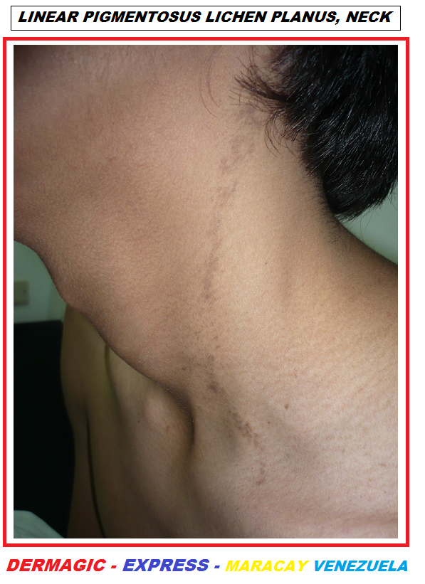
11.) Lineal.
12.)uco-membranoso (genital, esófago)
13.) Uñas.
14.) Oral.
15.) Penfigoide.
16.) Perforante.
17.) Pigmentoso.
18.) Plano Pilar.
19.) Ulcerativo
20.) Zosteriforme 
El oral tiene 6 tipos: Reticulado, atrófico, papular, en placa, erosivo y buloso. 
 
ENFERMEDADES ASOCIADAS:
-----------------------------------------------
Se han descrito numerosas enfermedades asociadas con LIQUEN PLANO entre ellas: 
 
A.) MALIGNAS:
----------------------
1.) Cáncer de estómago.
2.) Linfoma.
3.) Neuroblastoma.
4.) Adenoma de la pituitaria. 
5.) Fibrohistiocitoma.
6.) Paraproteinemia monoclonal por IGA Kappa.
7.) Craneofaringioma
8.) Malignidad pararenal.
9.) Sarcoma. 
 
B.) ENFERMEDADES GASTROINTESTINALES Y ENDOCRINAS.
----------------------------------------------------------------------------------------------
1.) Cirrosis biliar primaria.
2.) Hepatitis crónica activa. 
3.) Colitis Ulcerativa. 
4.) Diabetes mellitus.
5.) Anormalidad en el funcionamiento del hígado (enzimas)
 
C.) ENFERMEDADES AUTOINMUNES: 
----------------------------------------------------------
1.) Alopecia Areata.
2.) Dermatomiositis.
3.) Dermatitis Herpetiforme.
4.) Tiroiditis de Hashimoto.
5.) Queratoconjuntivitis seca y xerostomía.
6.) Morfea.
7.) Miastenia Gravis.
8.) Pénfigo foliáceo.
9.) Pénfigo Vulgar.
10.) Anemia perniciosa.
11.) Esclerosis sistémica.
12.) Timoma.
13.) Vitíligo.  
También el LIQUEN PLANO a sido asociado al uso de numerosas drogas y sustancias: 


LIQUEN PLANO BOCA

Lichen planus mouth, palate


DROGAS ASOCIADAS CON LIQUEN PLANO :

-------------------------------------------------------------

Numerosas drogas han estado involucradas en la aparición de LIQUEN PLANO, denominadas LIQUEN PLANO-LIKE reacciones, entre ellas: 


A.) ANTIHIPERTENSIVOS: Captopril, cloro tiazida, Enalapril, Hidroclorotiazida, Labetolol, Metildopa, practolol, propanolol, espironolactona. 
 

B.) ANTIBIÓTICOS: Demaclociclina, Etambutol, Griseofulvina, Ketoconazol, Levamisol, Acido para-amino-salicílico, estreptomicina, tetraciclina. 
 

C: AINES: (ANTIINFLAMATORIOS NO ESTEROIDEOS:
Naproxeno, Indometacina, Feclofenac, Diflunisal, Flurbiprofen, Benoxaprofen, Acido acetil salicílico. 


D: ANTIMALARICOS:
Cloroquina, Quinacrina, Quinidina, Quinina. 


E.) PSICOTROPICOS:
Carbamazepina, Levomepromazina, Metopromazina, 


F.) AGENTES REVELADORES DE FILMS:
4-Amino-N-dietil-analina sulfato (TTS), CD2, CD3, P-isopropilamino-difenilamina (IPPD) 


G.) SULFONILUREAS:
Clorpropamida, Tolazamida, Tolbutamida. 

 
H.) MISCELANEOS:

Alopurinol, anfenazole, arsénico, cinarizina, oro, meticran, Musk ambrette, penicilamina, probenecid, pirimetamina, mercaptopropionilglicina, piritioxina, Medios de radiocontraste, trihexifenidil, interferon-alpha-N1 


... y probablemente existan otras mas que no están en esta lista. 
Genéticamente se ha establecido una asociación de los antígenos HLA y EL LIQUEN PLANO, también el STRESS emocional es un factor desencadenante, el consumo de TABACO también se ha relacionado al mismo y las infecciones. 


LIQUEN PLANO CLÁSICO DEL PIE

 Liquen plano clásico del pie


ALTERNATIVAS TERAPÉUTICAS:

---------------------------------------------------
Se ha utilizado varias propuestas para el tratamiento del LIQUEN PLANO, entre ellas destacan: 
 

1.) RETINOIDES: Isotretinoina, Etretinato, Acitretin,
2.) GRISEOFULVINA.
3.) CICLOSPORINA A.
4.) ANTIBIÓTICOS: Penicilina, Isoniacida, Aureomicina, Trimetoprim-Sulfametoxazol, tetraciclina
5.) ANTIPARASITARIOS: Metronidazol, Levamisol.
6.) DAPSONA y TALIDOMIDA
7.) ANTIMALÁRICOS: cloroquina, hidroxicloroquina, quinina, quinacrina.
8.) RADIOTERAPIA.
9.) ANTIMETABOLITOS: Ciclofosfamida, metotrexato.
10.) CORTICOSTEROIDES: Clobetasol y acetonido de triamcinolona.
11.) MEDICACIÓN PSIQUIÁTRICA: Sulpiride.
12.) AZATIOPRINA.
13.) PUVA: psoralenos mas radiación UVA
14.) CIRUGÍA.
15.) VIEJAS TERAPIAS INCLUYEN: Mercurio sistémico, Acido nicotínico, Bismuto, vitaminas, calcio intravenoso, y arsenicales.

 LIQUEN PLANO DE LA LENGUA


liquen plano lengua


NUEVAS ALTERNATIVAS DE TRATAMIENTO:

------------------------------------------------------------
 
1.) DERIVADOS DE PLAQUETAS E INMUNOSUPRESIÓN.
2.) glycyrrhizin (LICORICE), HIERBA DE ORIGEN CHINO.
3.) TACROLIMUS Y PIMECROLIMUS TÓPICO
4.) HEPARINA (ENOXAPARIN)
5.) INTERFERON ALFA- 2b
6.) FOTOQUIMIOTERAPIA EXTRACORPOREA.
7.) MICOFENOLATO DE MOFETIL
8.) AMLEXANOX PASTA FORMULADA AL 5%
9.) LASER DE DIODO. (Terapia de Laser de bajo nivel)
10.) BARICITINIB (Inhibidor de la Janus Kinasa JAK 1 y 2)
11.) TOFACITINIB ( Inhibidor de la Janus Kinasa JAK 1 y 3)
12.) RUXOLITINIB (Inhibidor topico de la Janus Kinasa 1 y 2).
13. DEUCRACITINIB (Es un inhibidor selectivo de la tirosina quinasa 2 (TYK2, actualmente en estudio en el Liquen plano).
14. UPACITINIB (Inhibidor de la Janus Kinasa JAK 1).
 
Quizá una de las cosas mas importantes de esta revisión es LA GRAN ASOCIACIÓN QUE SE DESCRIBE actualmente ENTRE EL LIQUEN PLANO Y LA HEPATITIS CRÓNICA C Y B, y la VACUNACIÓN contra LA MISMA HEPATITIS B, o anormalidades en el funcionamiento hepático.
 

En base a todos estos hallazgos PODRÍAMOS CLASIFICAR  EL LIQUEN PLANO DENTRO DE 7 VARIANTES, en cuanto a su ORIGEN:
 
1.) ASOCIADO A ENFERMEDAD HEPÁTICA.
2.) ASOCIADO A OTRAS ENFERMEDADES NO HEPÁTICAS.
3.) INDUCIDO POR DROGAS Y CONTAMINANTES.
4.) 
IDIOPÁTICO.
5.) MARCADOR CUTÁNEO DE MALIGNIDAD.
6.) GENÉTICO (HLA ANTÍGENOS)
7.) ASOCIADO A VACUNACIÓN CONTRA HEPATITIS B

LIQUEN PLANO LINEAL: FRENTE A PUNTA DE NARIZ

Liquen plano lineal cara 

Probablemente Erasmus WILSON nunca pensó la gran relación que ha sido descubierta entre el LIQUEN PLANO  y ENFERMEDAD HEPÁTICA o anormalidades en el funcionamiento HEPÁTICO, y mas aun la aparición de esta enfermedad después de vacunación contra otra enfermedad (HEPATITIS B)... 

.. En estas 81 referencias los hechos en el adjunto: liquen plano clásico, lineal, boca y pecho.
 
En este enlace encontraras una ACTUALIZACIÓN DEL LIQUEN PLANO ORAL (2024).

Saludos a Todos.

Dr. José Lapenta R. 
Dr. José M. Lapenta.


EDITORIAL ENGLISH
===================
Hello friends of the net. DERMAGIC again with you. In this new review about the LICHEN PLANUS, disease described more than 150 years ago and that nowadays their treatment continues being an entire challenge for the dermatoligist of the new era.

ERASMUS WILSON was the first to use the term LICHEN PLANUS in 1869. The first variant of the disease was described by Kaposi in 1892, calling it  Lichen Ruber Pemphigoides ruber pemphigoids, and Frederic Wickham in 1895 described the WHITE STRIATIONS on the upper surface of the lesions.

Darier later described the histopathological characteristics of the disease in 1905.

After many years of scientific advancement, numerous factors have been clarified regarding LICHEN PLANUS. These factors are:

ETIOLOGY:


1. IMMUNOMEDIATION


Lichen planus is a chronic inflammatory disease currently considered of unknown etiology. It is caused by an
IMMUNE REACTION mediated by cytotoxic T lymphocytes that attack basal keratinocytes, causing damage and apoptosis in the skin and mucous membranes.

2. TRIGGERING FACTORS:
Several factors can act as triggers, including:

A. VIRAL INFECTIONS: the hepatitis C and hepatitis B viruses being the most important associations.

B.- MEDICINES:


- Antihypertensives: angiotensin-converting enzyme inhibitors (ACE inhibitors = enalapril) and beta-blockers: metoprolol, atenolol, propranolol, bisoprolol, carvedilol, labetalol, nebivolol).

- Antimalarials (chloroquine and hydroxychloroquine).

- Nonsteroidal anti-inflammatory drugs (NSAIDs): such as ibuprofen and naproxen, among others.

- Medications for type 2 diabetes: (sulfonylurea).

- Diuretics: thiazides.

- Penicillamine (used in rheumatic diseases).

These are the most frequently described, but in everyday experience, any medication can cause lichen planus. Lichen planus is often described in the literature as
LICHENOID DRUG ERUPTION, but histopathologically and clinically it is actually lichen planus.

C. CONTACT WITH ALLERGENS: Contact with dental allergens such as amalgam, and the developers used in X-ray films and photography.

3. GENETIC PREDISPOSITION:


There is a proven association of lichen planus with histocompatibility antigens, certain HLA haplotypes, class II molecules,
HLA-DR1, and the occurrence of familial cases, which demonstrates a genetic component in the susceptibility to this disease.

4.) ASSOCIATED AUTOIMMUNE DISEASES:

Lichen planus often coexists with other autoimmune diseases, including:

a. Alopecia areata.

b. Ulcerative colitis.

c. Chronic discoid lupus.

d. Reinforcing its immune nature.

e. Celiac disease.

f. Dermatomyositis.

g. Autoimmune thyroiditis (Hashimoto's).

h. Lichen sclerosus et atrophicus.

5.) ENVIRONMENTAL AND SYSTEMIC FACTORS:

Psychological stress, alterations in the microbiome (viruses, bacteria, and fungi that inhabit the human body), and chemical substances (already described) can also modify the immune response and trigger the onset of this pathology.

NOTE:

To date, no
SPECIFIC ORGANISM or LIVING AGENT has been described as the primary cause of lichen planus.

CLINICAL FEATURES:


It is characterized by flat, purplish, shiny lesions with polygonal papules that have whitish striations on their surface, called
WICKHAM'S STRIATIONS, named after its discoverer. The pruritus or itching is typically described in cycles (sometimes present, sometimes not).

The lesions can appear anywhere on the body, including mucous membranes and appendages such as nails and scalp.

On the oral mucosa, they appear as
whitish striations on the palate and the surface of the tongue. They are generally painful, including the genital region.

It can occur in both men and women, and usually in adults, but has also been described in adolescents and children.
THE DISEASE IS NOT CONTAGIOUS.

 
After many years the advance of the science had been able to decipher numerous events about the  LICHEN PLANUS, they are: 
 

Lichen planus pie 
 

CLINICAL TYPES OF LICHEN PLANUS:
-----------------------------------------------------
1.) Actinic.
2.) Annulare
3.) Bullous.
4.) Classic.
5.) Erythematosus.
6.) Exfoliative.
7.) familial
8.) Guttate.
9.) Hypertrophic.
10.) "invisible"
11.) Lineal.
12.) Muco-membranous (genital, esophagus)
13.) Nail.
14.) Oral.
15.) Penphigoides.
16.) Perforanting.
17.) Pigmentosus.
18.) Planopilaris.
19.) Ulcerative.
20.) Zosteriform.
The oral one has 6 types: Reticulated, atrophic, papular, plaquelike, erosive and bullous. 
 

ASSOCIATE DISEASES:
------------------------------------
Numerous illnesses associated with LICHEN PLANUS have been described, among them: 


A.) MALIGNANCIES:
-------------------------------
1.) Stomach Cancer.
2.) Lymphoma.
3.) Neuroblastoma.
4.) Adenoma of the pituitary.
5.) Fibrohistiocytoma.
6.) IGA Kappa monoclonal paraproteinemia.
7.) Craniopharyngioma
8.) Pararrenal malignancy.
9.) Sarcoma. 


liquen plano: lengua


B.) GASTROINTESTINAL AND ENDOCRINE DISEASE
-------------------------------------------------------------------------
1.) Primary biliar cirrhosis (PBC).
2.) Chronic active hepatitis (CAH).
3.) Ulcerative colitis.
4.) Diabetes mellitus.
5.) Abnormalities in the liver function (enzymes).

 C.) AUTOIMMUNE DISEASES:
 -------------------------------------------------------------------------
1.) Alopecia Areata. 
2.) Dermatomyositis. 
3.) Dermatitis Herpetiformis. 
4.) Hashimoto's Thyroiditis. 
5.) Keratoconjunctivitis sicca and xerostomia. 
6.) Morphea. 
7.) Myasthenia Gravis. 
8.) Penphigus foliaceus. 
9.) Penphigus Vulgaris. 
10.) Pernicious anemia. 
11.) systemic sclerosis. 
12.) Thymoma. 
13.) Vitiligo.
 
 
Liquen plano lineal.


Also the LICHEN PLANUS had been associated to the use of numerous drugs and substances: 


DRUG ASSOCIATED WITH LICHEN PLANUS:
--------------------------------------------
Numerous drugs have been involved in the appearance of LICHEN PLANUS, called LP-like reactions, among them: 


A.) ANTIHYPERTENSIVE: Captopril, chlorothiazide, Enalapril, Hydroclorothiazide, Labetolol, Methyldopa, practolol, propranolol, pironolactone. 
 

B.) ANTIBIOTICS: Demeclocycline, Ethambutol, Griseofulvin, Ketoconazole, Levamisole, Para-amino-salicylic acid, streptomycin, teracycline. 
 

C.) NON STEROIDAL ANTIINFLAMMATORY DRUGS:
Naproxen, Indomethacin, Feclofenac, Diflunisal, Flurbiprofen, Benoxaprofen, acetylsalicylic acid. 


D: ANTIMALARIALS:
Chloroquine, Quinacrine, Quinidine, Quinine, hydroxychloroquine.
       
 
E.) PSYCHOTROPIC /NEUROLOGIC:

Carbamazepine, Levomepromazine, Metopromazine, Olanzapine. 


F.) FILMS DEVELOPING AGENTS:
4-Amino-N-diethyl-analine sulfate (TTS), CD2, CD3, p-Isopropylamino-diphenylamine (IPPD). 


G.) SULFONYLUREAS:
Chlorpropamide, Tolazamide, Tolbutamide. 

 
H.) MISCELLANEOUS:

Allopurinol, anphenazole, arsenic, cinnarizine, gold, methycran, Musk ambrette, penicillamine, probenecid, phyrimethamine, mercaptopropionylglycine, pyrithioxin, radiocontrast media, trihexyphenidyl, interferon-alpha-N1. 


... and probably exist other but that are not in this list. 
Genetically an association of the HLA antigens and THE LICHEN PLANUS it has been observed, also the emotional STRESS is a causing factor, the consumption of TOBACCO has also been related to the same one, and the infections. 

 
THERAPEUTIC ALTERNATIVES:

-------------------------------------------
It has been used several proposals for the treatment of the LICHEN PLANUS, among them they highlight: 


1.) RETINOIDS: Isotretinoin, Etretinat, Acitretin, Temarotene.
2.) GRISEOFULVIN.
3.) CYCLOSPORINE A.
4.) ANTIBIOTICS: Penicillin, Isoniazid, Aureomycin, Trimethoprim-Sulfamethoxazole, tetracycline.
5.) ANTIPARASITE DRUGS: Metronidazole, Levamisole.
6.) DAPSONE and THALIDOMIDE
7.) ANTIMALARIALS: Phenytoin.
8.) RADIOTHERAPY.
9.) ANTIMETABOLITES: Cyclophosphamide, metotrexate.
10.) CORTICOSTEROIDS: Clobetasol and triamcinolone acetonide.
11.) PSYCHIATRIC MEDICATION: Sulpiride.
12.) AZATHIOPRINE.
13.) PUVA: psoralens plus UVA radiation.
14.) SURGERY.
15.) OLD THERAPIES INCLUDE: Systemic Mercury, nicotinic acid, Bismuth, vitamins, intravenous calcium, and arsenicals. 


OTHERS NEW ALTERNATIVES OF TREATMENT:
-----------------------------------------------------------------
 

1.) RECOMBINANT PLETELET-DERIVED GROWHT FACTOR AND INMUNOSUPRESION.
2.) glycyrrhizin (LICORICE), GRASS OF CHINESE ORIGIN.
3.) TACROLIMUS AND PIMECROLIMUS TOPIC.
4.) HEPARIN. (ENOXAPARIN)
5.) INTERFERON ALFA - 2b
6.) EXTRACORPOREAL PHOTOCHEMOTHERAPY.
7.) MOFETIL MYCOPHENOLATE 

8.) Amlexanox, formulated in a 5% paste 
9.) DIODE LASER (Low level laser therapy)
10.) BARICITINIB (Janus Kinase JAK 1 and 2 Inhibitor)
11.) TOFACITINIB (Janus Kinase JAK 1 and 3 Inhibitor)
12.) RUXOLITINIB (Topical Janus Kinase 1 and 2 Inhibitor).
13. DEUCRACITINIB (It is a selective inhibitor of tyrosine kinase 2 (TYK2, currently being studied in Lichen planus).
14. UPACITINIB (Janus Kinase JAK 1 Inhibitor).

Based on all these discoveries we could CLASSIFY THE LICHEN PLANUS on their origin IN SEVEN (7) VARIANTS. 


1.) ASSOCIATED TO HEPATIC DISEASES.
2.) ASSOCIATED TO OTHER NON HEPATIC DISEASES.
3.) INDUCED BY DRUGS AND POLLUTANTS.
4.) IDIOPATHIC.
5.) CUTANEOUS MARKER OF MALIGNANCY.
6.) GENETIC (HLA ANTIGENS)
7.) INDUCED BY HEPATITIS B VACCINE

Erasmus Wilson probably NEVER THOUGHT the great relationship that has been discovered between the LICHEN PLANUS AND HEPATIC  or ABNORMALITIES in the HEPATIC FUNCTION, And even more the appearance of the same after the vaccination against another disease (HEPATITIS B).

in these 81 references the facts ... in the attach clasical lichen planus, lineal, oral and chest manifestations !
 
In this link you will find an UPDATE ON ORAL LICHEN PLANUS (2024).

Greetings to all

Dr. Jose Lapenta
R.


===============================================================
REFERENCIAS BIBLIOGRÁFICAS
/ BIBLIOGRAPHICAL REFERENCES
===============================================================
A.- Updates In Therapeutics for Lichen Planus Pigmentosus (2022). 

B.- Review of Nail Lichen Planus: Epidemiology, Pathogenesis, Diagnosis, and Treatment (2021).
C.- Oral Lichen Planus: An Update on Diagnosis and Management (2024). D.- Treatment of Oral Erosive Lichen Planus With Upadacitinib (2022). E.- [Treatment of oral lichen planus-a review] (2025).
F.- Erosive Lichen Planus (2017). 
G.- Coexistence of oral lichen planus and vulvar lichen sclerosus (2025).
H.- Lichen Planus: What Is New in Diagnosis and Treatment? (2024).
I.- Hypertrophic Lichen Planus: An Up-to-Date Review and Differential Diagnosis (2024).
J.- Distinct Variations in Gene Expression and Cell Composition Across Lichen Planus Subtypes 2024).
K.- Successful Treatment of Erosive Lichen Planus With Tofacitinib: A Case Series and Review of the Literature 2024).
L.- JAK inhibitors in lichen planus: a review of pathogenesis and treatments (2022). 
M.- Successful treatment of childhood lichen planus with upadacitinib (2024). 
N.- Successful treatment of erosive lichen planus with Upadacitinib complicated by oral squamous cell carcinoma 2023).
O.- TYK2 inhibition with deucravacitinib ameliorates erosive oral lichen planus (2024).  
P.- Rapid response of lichen planus to baricitinib associated with suppression of cytotoxic CXCL13+CD8+ T cells (2024).
Q.- Recalcitrant multi-variant lichen planus successfully treated with oral baricitinib and topical ruxolitinib cream (2024).
R.- Mycophenolate mofetil and lichen planopilaris: systematic review and meta-analysis 2022).
S.- Assessment of 5% Amlexanox, 0.1% Triamcinolone Acetonide and 0.03% Tacrolimus in the Management of Oral Lichen Planus (2023).
T. Bidirectional Association between Lichen Planus and Hepatitis C-An Update Systematic Review and Meta-Analysis 2023).
W.- Survey of Medical Referral by Japanese Dentists for Patients With Hepatitis B, Hepatitis C, and Lichen Planus (2024). 
===============================================================

 1.) Lichen planus involving the esophagus.
2.) Hepatitis C virus infection prevalence in lichen planus: examination of lesional and normal skin of hepatitis C virus-infected patients with lichen planus for the presence of hepatitis C virus RNA.
3.) [Lichen planus and hepatitis C virus. Apropos of 5 new cases] TO: Lichen plan et virus de l'hepatite C. A propos de 5 nouveaux cas.
4.) Lichen planus occurring after hepatitis B vaccination: a new case.
5.) A case of oral lichen planus with chronic hepatitis C successfully treated by glycyrrhizin (LICORICE)
6.) Nail lichen planus in children: clinical features, response to treatment, and long-term follow-up.
7.) Azathioprine for the treatment of severe erosive oral and generalized lichen planus.
8.) Ulcerative lichen planus: a case responding to recombinant platelet-derived growth factor BB and immunosuppression.
9.) [Study on regulatory effect of composite taixian tablet on immune function of red blood cell in patients with oral lichen planus]
10.) Topical tacrolimus and pimecrolimus: future directions.
11.) Tacrolimus clinical studies for atopic dermatitis and other conditions.
12.) Low-dose low-molecular-weight heparin (enoxaparin) is beneficial inlichen planus: a preliminary report
13.) Low-dose low-molecular-weight heparin in lichen planus
14.) Management of recalcitrant ulcerative oral lichen planus with topical tacrolimus.
15.) Topical tacrolimus in the treatment of symptomatic oral lichen planus: a series of 13 patients.
16.) Mast cell degranulation and the role of T cell RANTES in oral lichen planus.
17.) Levamisole and/or Chinese medicinal herbs can modulate the serum level of squamous cell carcinoma associated antigen in patients with erosive oral lichen planus.
18.) Dramatic response to levamisole and low-dose prednisolone in 23 patients with oral lichen planus: a 6-year prospective follow-up study.
19.) Successful treatment of generalized lichen planus with recombinant interferon alfa-2b.
20.) [Prevalence of oral lichen planus and oral leukoplakia in 112 patients with oral squamous cell carcinoma]
21.) Dental metal allergy in patients with oral, cutaneous, and genital lichenoid reactions.
22.) [Cellular immune alterations in fifty-two patients with oral lichen planus.]
23.) Isolated lichen planus of the toe nails treated with oral prednisolone.
24.) Lichen planus-like eruption following autologous bone marrow transplantation for chronic myeloid leukaemia.
25.) Immune mechanisms in oral lichen planus.
26.) Cyclosporin A in the treatment of lichen planus.
27.) Oral metronidazole treatment of lichen planus.
28.)Idiopathic lichen planus: treatment with metronidazole.
29.) Intestinal amebiasis, lichen planus, and treatment with metronidazole.
30.) Urinary tract infection as a cause of lichen planus: metronidazole therapy.
31.) [Metronidazole treatment of the erosive ulcerative form of lichen ruber planus of the oral mucosa]
32.) Clinical and pathological characteristics of oral lichen planus in hepatitis C-positive and -negative patients.
33.) High prevalence of anticardiolipin antibodies in patients with HCV-associated oral lichen planus.
34.) The clinical features, malignant potential, and systemic associations of oral lichen planus: a study of 723 patients.
35.) Management of oral lichen planus.
36.) Lichen planus occurring after hepatitis B vaccination: a new case.
37.) TT virus detection in oral lichen planus lesions.
38.) Lichenoid eruption following hepatitis B vaccination: first North American case report.
39.) Increased frequency of HLA-DR6 allele in Italian patients with hepatitis C virus-associated oral lichen planus.
40.) Extrahepatic manifestations of chronic viral hepatitis.
41.) Prevalence of hepatitis C virus in patients with lichen planus of the oral cavity and chronic liver disease.
42.) Histopathological and immunohistochemical study of oral lichen planus-associated HCV infection.
43.) Previous tuberculosis, hepatitis C virus and lichen planus. A report of 10 cases, a causal or casual link?
44.) [Skin diseases and hepatitis virus C infection]
45.) [Extrahepatic manifestations of hepatitis C virus infection]
46.) Detection of hepatitis C virus RNA in oral lichen planus and oral cancer tissues.
46.) Oral lichenoid lesions after hepatitis B vaccination.
47.) [The extrahepatic manifestations in hepatitis C virus (HCV) infection]
48.) Association of HLA-te22 antigen with anti-nuclear antibodies in Chinese patients with erosive oral lichen planus.
49.) Treatment of autoimmune and extra-hepatic manifestations of HCV infection.
50.) Lichen planus, erythema nodosum, and erythema multiforme in a patient with chronic hepatitis C.
51.) [Clinical considerations and statistical analysis on 100 patients with oral lichen planus]
52.) Lichen planus in children: a possible complication of hepatitis B vaccines.
53.) Lichen planus actinicus treated with acitretin and topical corticosteroids.
54.) Alendronate-induced lichen planus.
55.) Hepatitis C virus and lichen planus in Nigerians: any relationship?
56.) Helicobacter pylori Infection in Skin Diseases: A Critical Appraisal.
57.) Presence of lichen planus during a course of interferon alpha-2a therapy for a viral chronic C hepatitis.
58.) Lichen planus-like eruption following autologous bone marrow transplantation for chronic myeloid leukaemia.
59.) [Clinical evaluation in oral lichen planus with chronic hepatitis C: the role of interferon treatment]
60.) Oral lichen planus induced by interferon-alpha-N1 in a patient with hepatitis C.
61.) Treatment of lichen planus. An evidence-based medicine analysis of efficacy.
62.) Successful Treatment of Resistant Hypertrophic and Bullous Lichen Planus With Mycophenolate Mofetil
63.) Liver abnormalities in patient with lichen planus.
64.) Lichen planus.
65.) Evaluation of Hepatitis B Vaccination among Lichen Planus Patients.
66.) Lichen planus associated with hepatitis C virus: no viral transcripts are found in the lichen planus, and effective therapy for hepatitis C virus does not clear lichen planus.
67.) Lichen planus secondary to hepatitis B vaccination.
68.) A clinical evaluation of the efficacy of photodynamic therapy in the treatment of erosive oral lichen planus: A case series.
69.) Possible alternative therapies for oral lichen planus cases refractory to steroid therapies.
70). Novel therapies for oral lichen planus.
71.) The effect of diode laser and topical steroid on serum level of TNF-alpha in oral lichen planus patients.

===============================================================

Si Te ha gustado, Compartelo